Gharbi Classification Usg - The Surgical Management Of Hydatid Cyst Of The Liver What Is New Intechopen / Type i appears cystic and unilocular.. Patient had a maximum flow rate of 11.3 ml/s and an average flow rate of 7.4 ml/s in uroflowmetry. Gharbi classification is shown in table i. In order to assess the relation of the cyst with the biliary tract, the relevant ducts were visualized following the irrigation of the cavity via isotonic after the drainage of the cyst had been achieved. Type ultrasound appearance i pure fluid collection ii fluid collection with a split wall iii fluid collection with septa iv heterogeneous echo pattern v reflecting walls type v cysts determined by ultrasound to be calcified and have been assumed to be dead cysts and do not. 3.2 gharbi classification in usg.
radiology 139 (1981) 459 which has been widely used, but in modified forms, since its publication. Even though ultrasonographic evaluation is known to be of Gharbi classification of these patients who were cured revealed that six patients had gharbi iii, four had gharbi ii, and two had gharbi i cysts. Mechanical suction through wide bore catheters for nonsurgical management of gharbi type iii hepatic hydatid cysts: Type ultrasound appearance i pure fluid collection ii fluid collection with a split wall iii fluid collection with septa iv heterogeneous echo pattern v reflecting walls type v cysts determined by ultrasound to be calcified and have been assumed to be dead cysts and do not.
Mechanical Suction Through Wide Bore Catheters For Nonsurgical Management Of Gharbi Type Iii Hepatic Hydatid Cysts from www.tropicalgastro.com 3.2 gharbi classification in usg type 1: Patient was again followed up at 6 months and was doing well. There are more than 15 classification schemes for liver hydatid cysts.the liver is the most common site of hydatid disease involvement, and most cysts are located in the right lobe.a cl (cystic lesion) type has been added in the who classification that was not included in gharbi's classification, for those cysts whose parasitic nature cannot. In our series, we included 18 cysts of gharbi type iii, 9 of type ii, and 8 of type i. The gharbi classification system was used to stage the hydatid disease. Suyash mohan, 1 shobhit k garg, 2 manoj kathuria, 3 sanjay saran baijal 2 department of radiology, 1 university of michigan health system, The overall success rate of albendazole therapy was 18% (12/65) in the study. Granulosus cyst fluid samples from naturally infected pigs.
In our series, we included 18 cysts of gharbi type iii, 9 of type ii, and 8 of type i.
After 3 months usg was done which showed no liver hydatid cyst. Type ii is a fluid filled with a floating membrane (the water lily sign). The classification proposed by gharbi et al., for liver hydatid disease based on usg appearance, can be adopted for other locations also. Touch device users can explore by touch or with swipe gestures. The gharbi classification system was used to stage the hydatid disease. Patient had a maximum flow rate of 11.3 ml/s and an average flow rate of 7.4 ml/s in uroflowmetry. 10 who classification is almost the same as gharbi's, with gharbi type ii corresponding to ce 3a of. Hydatid cyst demonstrates a variety of imaging features, varying according to growth stage, associated complications, and affected tissue. There are more than 15 classification schemes for liver hydatid cysts.the liver is the most common site of hydatid disease involvement, and most cysts are located in the right lobe.a cl (cystic lesion) type has been added in the who classification that was not included in gharbi's classification, for those cysts whose parasitic nature cannot. This classification was proposed by the who in 2001 and, at the time of writing (july 2016), remains the most widely used classification for hepatic hydatid cysts. 9 hassen gharbi classified hepatic hydatid into five types based on sonographic appearance. The decomposition of the germinative membrane and the pericyst. Patient was again followed up at 6 months and was doing well.
Granulosus cyst fluid samples from naturally infected pigs. Patient was again followed up at 6 months and was doing well. After 3 months usg was done which showed no liver hydatid cyst. There was only one case of type iv and no cases of type v. There are more than 15 classification schemes for liver hydatid cysts.the liver is the most common site of hydatid disease involvement, and most cysts are located in the right lobe.a cl (cystic lesion) type has been added in the who classification that was not included in gharbi's classification, for those cysts whose parasitic nature cannot.
Hydatid Diseases from image.slidesharecdn.com There are more than 15 classification schemes for liver hydatid cysts.the liver is the most common site of hydatid disease involvement, and most cysts are located in the right lobe.a cl (cystic lesion) type has been added in the who classification that was not included in gharbi's classification, for those cysts whose parasitic nature cannot. In our series, we included 18 cysts of gharbi type iii, 9 of type ii, and 8 of type i. 3.2 gharbi classification in usg. Only for gharbi classification type 1 and 2 (table). Usg appearance of hydatid cyst. 10 who classification is almost the same as gharbi's, with gharbi type ii corresponding to ce 3a of. Type ultrasound appearance i pure fluid collection ii fluid collection with a split wall iii fluid collection with septa iv heterogeneous echo pattern v reflecting walls type v cysts determined by ultrasound to be calcified and have been assumed to be dead cysts and do not. Ultrasonographic appearance of a calcified hydatid cyst.
Efficacy is 100%, but the treatment should be preserved only for gharbi classification type 1 and 2 (table).
Ultrasonography may sometimes be insufficient for the differential diagnosis of the lesion seen in the images. Usg appearance of hydatid cyst. Calcified or partially calcified lesion (inactive cyst) The classification proposed by gharbi et al., for liver hydatid disease based on usg appearance, can be adopted for other locations also. radiology 139 (1981) 459 which has been widely used, but in modified forms, since its publication. Hydatid disease is a worldwide zoonosis produced by the larval stage of the echinococcus tapeworm (, fig 1).the two main types of hydatid disease are caused by e granulosus and e multilocularis.the former is commonly seen in the great grazing regions of the world—particularly the mediterranean region, africa, south america, the middle east, australia, and new zealand—and is. Discover (and save!) your own pins on pinterest After 3 months usg was done which showed no liver hydatid cyst. The classification proposed follows that of the first classification developed by gharbi et al. The decomposition of the germinative membrane and the pericyst. In cases of clinical suspicion and presence of predictive factors, further studies can be planned such as mrcp or endoscopic retrograde cholangiopancreatography (ercp). Cyst < 6 cm cyst > 6 cm growth of cyst > 2 cm /peryear growth of cyst < 2 cm /peryear: The decomposition of the germinative membrane and the pericyst.
Efficacy is 100%, but the treatment should be preserved only for gharbi classification type 1 and 2 (table). The mean size of the cysts In our series, we included 18 cysts of gharbi type iii, 9 of type ii, and 8 of type i. Ct gives more detailed information about the localization and size of the cyst, and its sensitivity is 100% (3,11). Gharbi classification on ultrasonographic features of hydatid cyst3 10.
The Gharbi Classification For Hydatid Cysts Download Table from www.researchgate.net 9 hassen gharbi classified hepatic hydatid into five types based on sonographic appearance. Ultrasonography may sometimes be insufficient for the differential diagnosis of the lesion seen in the images. Ultrasonography (usg) and computed tomography (ct) are both valuable imaging methods for the diagnosis of liver hydatid disease. Gharbi classification is shown in table i. Discover (and save!) your own pins on pinterest Touch device users can explore by touch or with swipe gestures. radiology 139 (1981) 459 which has been widely used, but in modified forms, since its publication. The decomposition of the germinative membrane and the pericyst.
3.2 gharbi classification in usg.
The mean size of the cysts According to gharbi's classification, three cases (21.4 %) of the unusually located hydatid cysts were type i, two (14.3 %) type ii, and eight (57.1 %) type iii. The classification proposed by gharbi et al., for liver hydatid disease based on usg appearance, can be adopted for other locations also. 9 hassen gharbi classified hepatic hydatid into five types based on sonographic appearance. Spleen and kidneys are the organs where hydatid disease is most frequently observed after the liver and lung. 3.2 gharbi classification in usg type 1: Type ultrasound appearance i pure fluid collection ii fluid collection with a split wall iii fluid collection with septa iv heterogeneous echo pattern v reflecting walls type v cysts determined by ultrasound to be calcified and have been assumed to be dead cysts and do not. The overall success rate of albendazole therapy was 18% (12/65) in the study. Ultrasonography (usg) and computed tomography (ct) are both valuable imaging methods for the diagnosis of liver hydatid disease. Transrectal usg revealed a homogeneous cystic mass not related to the bladder. The gharbi ultrasound classification consists of five stages 4: The classification proposed follows that of the first classification developed by gharbi et al. Only for gharbi classification type 1 and 2 (table).
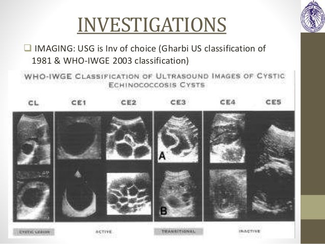
Gharbi classification of these patients who were cured revealed that six patients had gharbi iii, four had gharbi ii, and two had gharbi i cysts gharbi. The 2001 world health organization (who) classification of hepatic hydatid cysts is used to assess the stage of hepatic hydatid cysts on ultrasound and is useful in deciding the appropriate management depending on the stage of the cyst.


0 Komentar